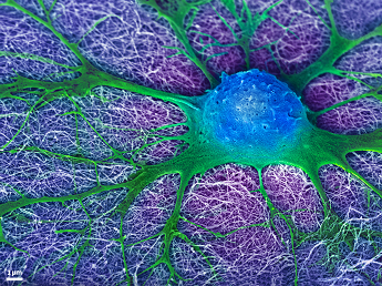
Caption:
False color image of an embryonic neural cell (blue/green) derived from the mouse spinal cord and cultured on a synthetic nanofiber gel (purple). The prolongations of the cell (green) are extending throughout the nanofibers to re-establish a neural network with other cells. These nanofibers (~10nm in diameter) were specifically designed to encourage neural outgrowth by mimicking the environment of spinal cord tissue.
Zaida Alvarez Pinto
Mark McClendon
Advisor: Samuel Stupp
Simpson Querrey Institute for BioNanotechnology
Northwestern University, Evanston, IL 60208
Laboratory website: http://stupp.northwestern.edu/group/index.html
Technique: Scanning Electron Microscopy, false color added with Photoshop
Description:
Spinal cord injuries often lead to lifelong disabilities and the key to functional recovery is reconnecting the neuronal pathways that were damaged. One solution is to induce neural stem cells in the spinal cord to regenerate the cellular extensions while discouraging scar tissue formation that would otherwise block the reconnections from occurring. Shown in this image is a mouse neural cell growing on a synthetic nanofiber gel that has been designed to mimic the native environment of the spinal cord tissue. A local injection of these nanofibers following spinal cord injuries in mice was able to reduce glial scar formation and restore hind leg function with only one treatment.
More Info:
J. Neurosci., 28(14), (2008) 3814-3823
Journal of Neuroscience Research 88(14), (2010) 3161-3170
Biomaterials 43(20), (2013) 4749-4757
Biomaterials 35(1), (2014) 185-195
Biomaterials 35(31), (2014) 8780-8790
Funding Source: National Institute of Health (NIH), Institute: National Institute of Biomedical Imaging and Bioengineering (NIBIB), Project #5R01EB003806-10
Beatriu de Pinós Fellowship (AGAUR)