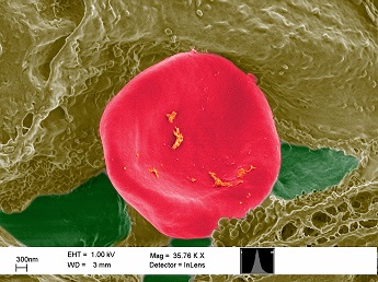
Caption:
A single red blood cell (erythrocyte) is visible in all its glory escaping out of a blood vessel in a porcine skin sample exposed to cerium oxide nanoparticles.
Sahil Tahiliani, Masters Student & Abhishek Gottipati, PhD Student
Advisor: Sara Brenner MD, MPH.
SUNY Polytechnic Institute Colleges of Nanoscale Science & Engineering
Nanobioscience Constellation
Albany, NY
Laboratory website: https://sunypoly.edu/research/team-brenner/
Technique: A thin section of porcine skin exposed to cerium oxide nanoparticles was imaged with a scanning electron microscope.
Description:
As nanomaterials are increasingly incorporated into manufacturing processes and consumer products, the assessment of potential occupational exposure to these materials is crucial to determine safe occupational practices. Although the skin has very effective protective properties, some nanoparticles may cross the skin barrier and produce different potentially inflammatory responses. This sample corresponds to pig skin topically exposed to cerium oxide nanoparticles contained in a solution that is regularly used in a polishing process during production of computer chips. Identification, location, and penetration of these nanoparticles is assessed by imaging these samples in high-resolution electron microscopes, such as a scanning electron microscope (SEM). These nanoparticles penetrate through into the different layers of skin but very rarely through the blood vessels. This is why there is the special focus on finding a ruptured blood vessel and a single RBC was imaged and false colored to see life in a different light.
Funding Source: NanoHealth & Safety Center, New York State (awarded to P.I.)