Caption:
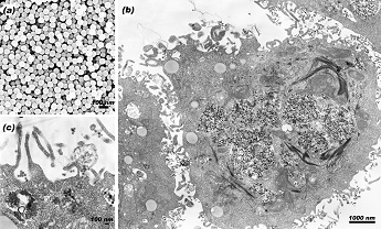
Scanning and transmission electron micrographs of silica nanoparticles (SNPs) with 60 nm diameters (a), lung cell exposed to high dose of 60 nm SNPs, showing fragmented nuclei and destruction of the cytosol (b), and 60 nm SNPs at the nano-bio interface and within the cytoplasm after cellular uptake (c).
In-Yong Kim
Department of Electrical and Computer Engineering
University of Illinois at Urbana-Champaign
Urbana, IL, USA
Laboratory website: http://tfcp.ece.illinois.edu/index.htm
Technique: Scanning and transmission electron microscopy
Description:
Nanoparticles are powerful tools in nanomedicine. This work indicates that cytotoxicity of nanoparticles involves changes in membrane integrity induced by cellular uptake. Silica nanoparticles (SNPs) administered as a drug delivery vehicle should be selected based on the interactions between the particles and the target organ or tissue, e.g. whether the SNPs preserve viability or induce cytotoxic, genotoxic, and epigenetic effects at target sites, and cytotoxicity should be assessed for multiple cells in order to get a full picture of the interactions between nanomaterials and biological tissues. In particular, 60 nm SNPs affected cells differently than nanoparticles of other sizes, most likely due to enhanced uptake; therefore, 60 nm particles may be ideal for biomedical applications requiring cellular uptake, but fatal at high doses. The picture is a warning that nanoparticles should be considered a double-edged sword for the safe and ideal design of multi-functionalized SNPs for biomedical applications.
Funding Source: This work was supported in part by the University of Illinois Center for Nanoscale Science and Technology (CNST), the National Cancer Institute-funded Siteman Center for Cancer Nanotechnology Excellence at Illinois, and Trionix Research Laboratory, Inc