Caption:
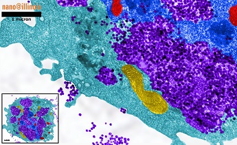
False color image of 60 nm silica nanoparticles (SNPs) in a cell. Inset shows the entire cell. Although the cell is dying and many cellular structures are destroyed, particles are still actively being taken in. Purple – SNPs; yellow – mitochondria; blue – nucleus; red – golgi. Scale bar = 1 µm.
Inyong Kima, Elizabeth Sawickib, Hyungsoo Choia, Kevin Kima,b
Advisor:
aDepartment of Electrical and Computer Engineering
University of Illinois at Urbana-Champaign
bDepartment of Bioengineering
University of Illinois at Urbana-Champaign
Technique: Transmission electron microscopy
Description:
We are investigating the toxicity of different sizes and doses of silica nanoparticles (SNPs) on various cell types. So far, we have found that a cell’s response to SNPs depends on cell type; some cell types are easily damaged by SNPs while others are more robust. By imaging cells treated with SNPs, like the one shown here, we discovered that a diameter of about 60 nm is the optimum size for cellular uptake. Cells will engulf large quantities of 60 nm SNPs even if the end result is cell death. We also discovered that different sizes of SNPs enter different parts of the cell: large particles are often found in mitochondria while medium particles are typically enclosed in vesicles and small particles can be found free inside the cytoplasm. Understanding the interactions between cell type and nanoparticle size is important for designing nanotechnologies that are safe for human use.