Caption:
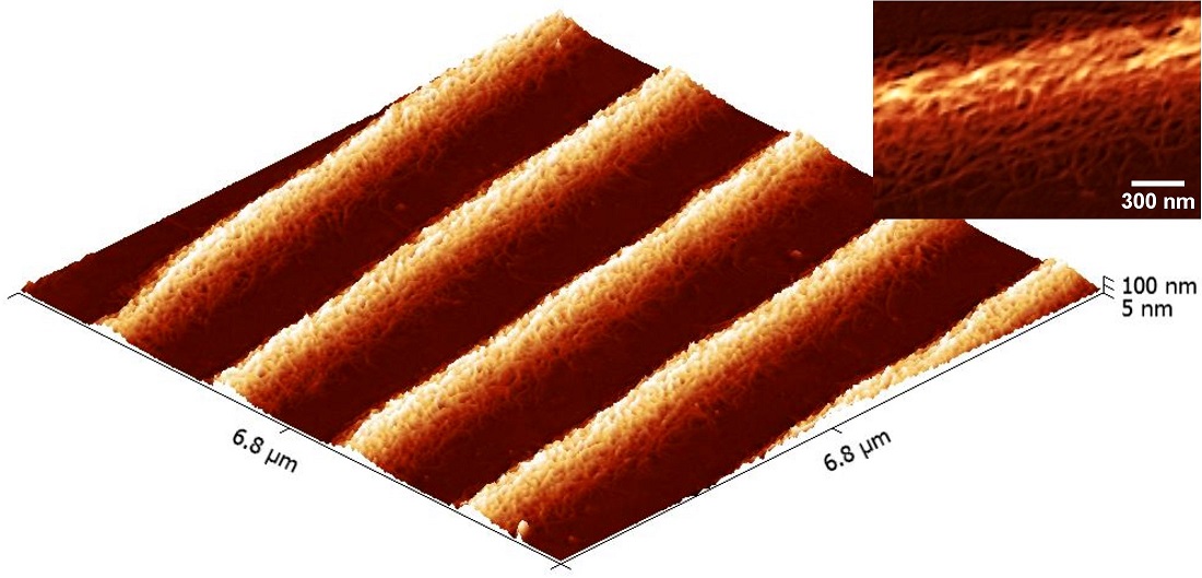
3-D topographical image of a mixture of chitin and collagen nanofibers that have been molded into micron-wide, parallel tracks. The inset is a magnified top-down image showing the texture of the nanofibers which are 10-20 nm in diameter.
Kimberly Riddick
Advisor: Dr. Albert Hung
University of North Carolina Greensboro
Joint School of Nanoscience and Nanoengineering
Department of Nanoengineering
Laboratory website: http://jsnn.ncat.uncg.edu
Technique: The image was acquired using a Keysight Technologies 5600LS AFM and Budget Sensors SiNi silicon nitride cantilever tips (0.06 N/m force constant). Image processing was performed using Gwyddion open source software and ImageJ (NIH).
Description:
One goal of biomedical research is to find ways of replacing diseased or damaged organs or tissue in the body, either by encouraging the body to regrow them on its own or by growing viable replacements in the lab artificially, eliminating the need for donors. To do this, cells might be seeded on a “scaffold,” a structural skeleton made of non-toxic, biodegradable material that encourages the cells to grow with the proper shape and organization as real tissue. Chitin and collagen are both biological materials obtained naturally that assemble into nanoscale fibers that, if properly aligned, might guide cell growth and orientation. In this sample, a mixture of chitin and collagen was molded into micron-wide tracks using a rubber mold with channels. The goal of this research is to study the fundamental science behind the assembly and alignment of these nanofibers for the purpose of developing complex tissue scaffolds.
Funding Source: This work was supported through generous support of Dr. James Ryan, the Joint School of Nanoscience and Nanoengineering, and the State of North Carolina.