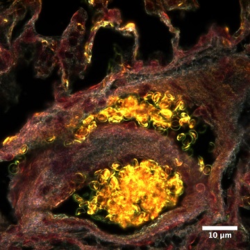
Caption:
Region of a lung with bright yellow blood vessel clusters containing oval discs of red blood cells, as shown by darkfield microscopy at a 100x objective lens magnification.
Sahil Tahiliani
Advisor: Dr. Sara Brenner
SUNY Polytechnic Institute Colleges of Nanoscale Science & Engineering
Nanobioscience Constellation
Albany, NY
Laboratory website: https://sunypoly.edu/research/team-brenner/
Technique:
Rat lung tissue was stained with hematoxylin and eosin (H&E), which are standard histological stains, for optical darkfield microscopy. This image was taken using a CytoViva enhanced darkfield microscope. Red blood cells appear as gold discs in blood vessels in the lung.
Description:
A rat model of inhalation exposure is used to study nanoparticle distribution throughout the body and any potential resulting adverse health effects. Following inhalation exposure to engineered metal oxide nanoparticles, inflammation was detected in rat lung. The red blood cells, with their oval biconcave disc shapes, appear as clusters bound together in the vessels. The stain and enhanced darkfield microscope cause the yellow color of the cells, which are the lifeline of the connective tissue.
Funding Source: The National Institute for Occupational Safety and Health and the NanoHealth & Safety Center, New York State