Caption:
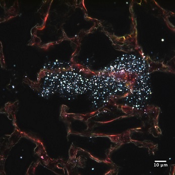
100x magnification of a collection of metal oxide nanoparticles within lung tissue
Leonardo Bezarra
Advisor: Dr. Sara Brenner
SUNY Polytechnic Institute Colleges of Nanoscale Science & Engineering
Nanobioscience Constellation
Albany, NY
Laboratory website: https://sunypoly.edu/research/team-brenner/
Technique:
Optical enhanced darkfield microscopy was performed on the lung tissue of a rat following inhalation exposed to cerium oxide nanoparticles. Tissues were stained with standard hematoxylin and eosin (H&E) histological stains and imaged using a CytoViva enhanced darkfield microscope. In this image, nanoparticles can be seen scattered throughout lung tissue.
Description:
The rapid growth and projected acceleration of nanotechnology creates urgency in understanding, predicting, and managing the health risks associated with workplace exposure to engineered nanomaterials. People may be unintentionally exposed to nanomaterials by breathing them in or touching them. Our group seeks to understand how the body reacts to the metal oxide nanoparticles used during semiconductor manufacturing. By using rats as a model of inhalation exposure, we hope to discover where the nanoparticles accumulate within the body and better understand associated immune responses.
Funding Source: The National Institute for Occupational Safety and Health and the NanoHealth & Safety Center, New York State