Caption:
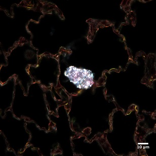
100x magnification of a collection of bright nanoparticles caught within rat lung tissue
Ahlam Abuawad
Advisor: Dr. Sara Brenner
SUNY Polytechnic Institute Colleges of Nanoscale Science & Engineering
Nanobioscience Constellation
Albany, NY
Laboratory website: https://sunypoly.edu/research/team-brenner/
Technique: Optical enhanced darkfield microscopy of rat tissues exposed to slurry via inhalation. Tissues were stained with standard Hematoxylin and Eosin (H&E) stains and imaged using a cytoviva microscope.
Description:
Workers in the semiconductor industry can be exposed to many different substances, but little is known about the health effects of this exposure, however. Our group seeks to understand how the body reacts to exposure to the metal nanoparticles used during a process called Chemical Mechanical Planarization, and where these compounds may accumulate within the body. By using rats as a model, we hope to discover how the body deals with the nanoparticles, where they accumulate within the body and any immune response as a result.
Funding Source: The National Institute for Occupational Safety and Health and the NanoHealth & Safety Center, New York State