Caption:
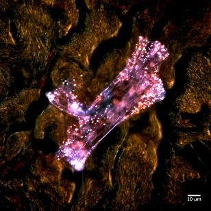
Metal oxide nanoparticles bright up over the middle layer of skin tissue with darkfield microscopy at a 100x magnification.
María del Pilar Sosa Peña
Advisor: Dr. Sara Brenner
SUNY Polytechnic Institute Colleges of Nanoscale Science & Engineering
Nanobioscience Constellation
Albany, NY
Laboratory website: https://sunypoly.edu/research/team-brenner/
Technique:
The pigskin sample was exposed to silica nanoparticles and then stained with hematoxylin and eosin, for optical darkfield microscopy. This image was taken using enhanced darkfield microscopy (EDFM) with a CytoViva microscope.
Description:
Studying the effect of metal oxide nanoparticles on tissue is necessary to understanding the potential risks of exposure. This is crucial for determining safe practices which will protect workers, particularly in the semiconductor industry. Skin is very good at protecting the body. However, nanoparticles may cross the skin barrier and cause different responses. This image is of a sample of pig skin topically exposed to a nanoparticle solution which is regularly used in the production of semiconductors. Identifying, locating, and determining how deeply the nanoparticles penetrated the skin was done using a CytoViva enhanced darkfield microscope.
Funding Source: The National Institute for Occupational Safety and Health and the NanoHealth & Safety Center, New York State


