Caption:
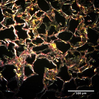
Region of a lung with bright yellow red blood cell clusters and bright white spots of metal oxide nanoparticles can be seen with darkfield microscopy at a 40x objective lens magnification.
Sahil Tahiliani
Advisor: Dr. Sara Brenner
SUNY Polytechnic Institute Colleges of Nanoscale Science & Engineering
Nanobioscience Constellation
Albany, NY
Laboratory website: https://sunypoly.edu/research/team-brenner/
Technique:
The rat lung tissue was stained with hematoxylin and eosin (H&E), for optical darkfield microscopy. This image was taken using enhanced darkfield microscope (EDFM) by CytoViva.
Description:
The goal of the study is to determine the toxicity of metal oxide nanoparticles used in semiconductor manufacturing in rats following inhalation exposure. This particular image shows the architecture of the alveolar region of the left lung of a rat exposed to silica nanoparticles via inhalation. Inflammation was detected in the rat lung following inhalation exposure to engineered metal oxide nanoparticles.
Funding Source: The National Institute for Occupational Safety and Health and the NanoHealth & Safety Center, New York State